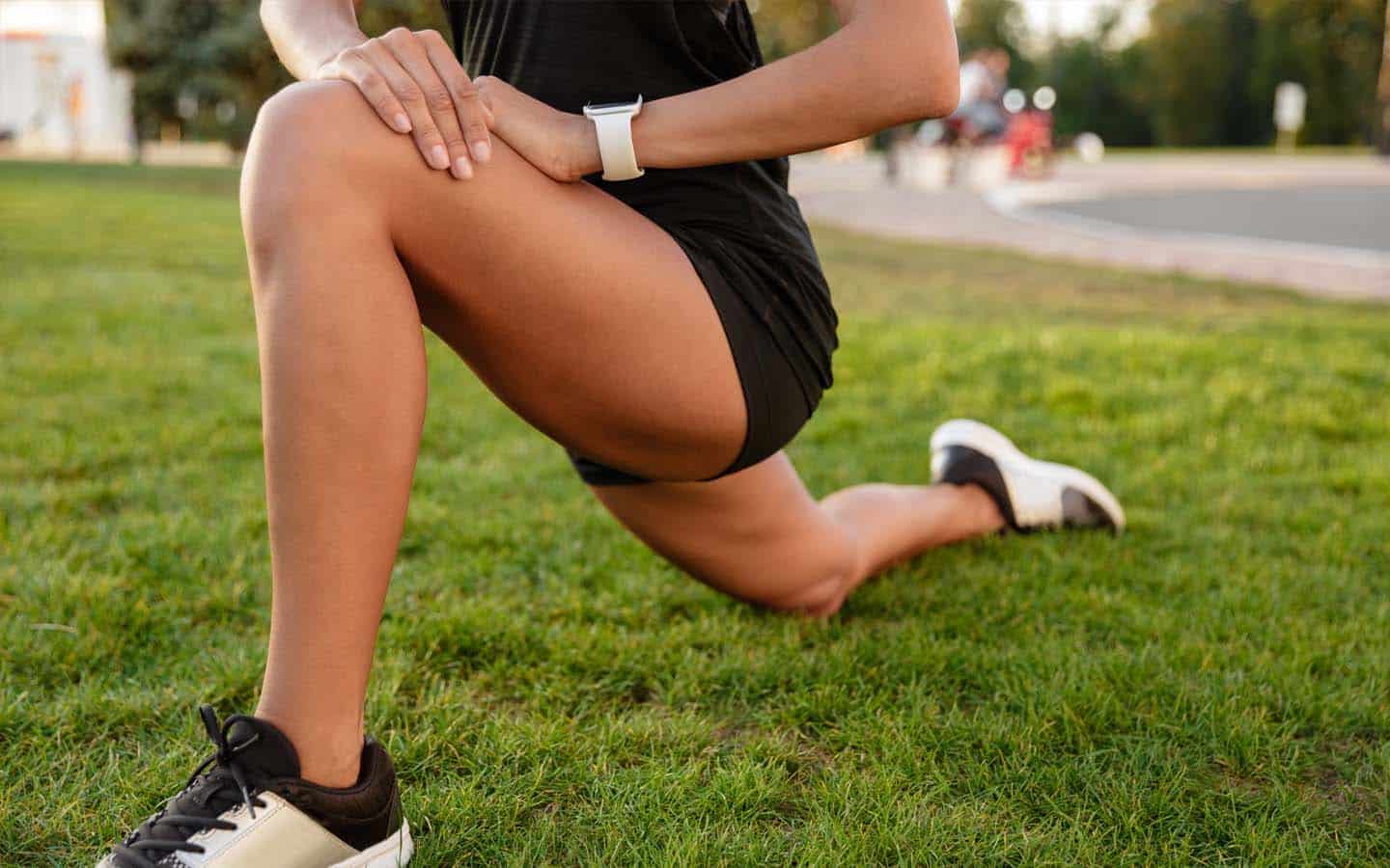
Anatomy Corner: Knee
The knee joint is the largest joint in the body. It consists of the patella (kneecap), femur (thigh bone), and tibia (shin bone). There is another bone next to the tibia called the fibula that is not part of the knee joint, but serves as an attachment point for one of the knee ligaments. The components of the knee joint are the patellofemoral joint and the tibiofemoral joint. The knee is primarily a hinge joint, but there is also some rotation.
The femur is the largest bone in the body and is very strong. The patella is the largest sesamoid bone (bone embedded in a tendon or muscle) in the body and is an attachment for the quadriceps tendon and the patellar tendon.
Stabilizing Ligaments of the Knee
The collateral ligaments are on the sides of the knee (medial collateral ligament and lateral collateral ligament) and provide stability side to side. The cruciate ligaments inside the knee – anterior cruciate ligament (ACL) and posterior cruciate ligament (PCL). The ACL stops the tibia from sliding forward on the femur. The PCL stops the femur from sliding forward on the tibia.
The menisci are fibrocartilaginous structures that are crescent shaped – one on the medial side and one on the lateral side. These cover most of the top of the tibia. They serve as shock absorbers and static stabilizers for the knee. Meniscus injuries are common, and can happen with a pivot on a planted foot. Meniscal injuries can also occur because of degenerative changes (with wear and tear).
The femur and the tibia are covered in articular cartilage, a smooth protective surface. Arthritis occurs with the degeneration of this surface.
The joint is contained by a joint capsule consisting of an inner synovial layer (secretes joint lubricating fluid) and an outer ligamentous layer.
There are two main muscle groups that span the knee joint. The quadriceps include the rectus femoris, vastus lateralis, vastus intermedius, and vastus medialis. These muscles act to extend or straighten the knee. The hamstrings include the biceps femoris, semitendinosus, and semimembranosus. These muscles act to flex or bend the knee.
Hip musculature plays a role in knee positioning, especially the abductors and external rotators.
By Eilish O’Sullivan

